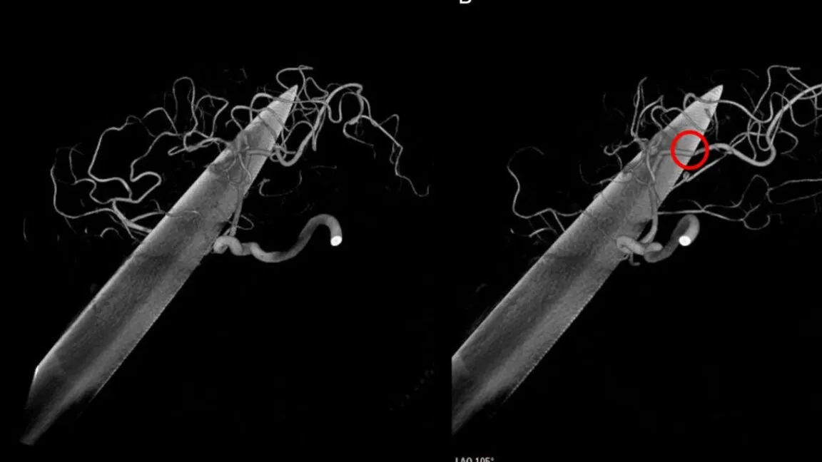
A penetrating brain injury involving a knife through the eye socket resulted in a remarkable recovery for a man in his 20s in the UK. Despite a 15 mm-wide blade traversing his right eye and through the left frontal and temporal lobes, the patient remained conscious and neurologically intact following emergency surgery. According to a report in BMJ Case Reports (2026), the patient’s survival and preservation of function were attributed to the specific trajectory of the blade, which avoided critical vascular and subcortical structures.
Clinical presentation and initial assessment
The patient arrived at a UK hospital with the hilt of a knife protruding from his face, having sustained a transorbital injury that penetrated the skull. Although the injury appeared catastrophic, the man was alive and conscious upon arrival, though medical staff noted he was in a state of agitation. His neurological status was measured using the Glasgow Coma Scale, where he scored a 12 (E3 V4 M5), classifying the injury as "moderate" rather than "mild."
His clinical presentation included several specific neurological markers:
- Eye response (E3): He could open his left eye when prompted but otherwise kept it closed.
- Verbal response (V4): He was able to talk and answer questions, though he exhibited confusion.
- Motor response (M5): He showed intentional movement, such as pulling away when pressure was applied to his body.
Due to his agitated and confused state, doctors expressed concern that the patient might attempt to remove the knife himself. To prevent self-inflicted trauma, medical teams immediately sedated and intubated him, while simultaneously initiating prophylactic treatments for bacterial infections and tetanus.
The trajectory of the blade through the brain
Advanced imaging, including CT angiographic reconstructions, revealed that the 15 mm-wide blade had traversed a complex path through the cranial compartment. The blade entered through the right orbit, completely passing through the eyeball and the eye socket before entering the brain. It moved through the left frontal and temporal lobes, following a trajectory that narrowly missed several vital neurological structures.
The precision of the path was a deciding factor in the patient's survival. The blade passed in front of the corpus callosum—the bundle of nerve fibers connecting the brain's two hemispheres—without causing apparent injury to it. Additionally, the knife missed the left internal capsule, another critical bundle of nerve fibers essential for motor and sensory functions. While the blade was located near the left internal carotid artery, imaging indicated that this major blood vessel had not been damaged.
Vascular implications and surgical findings
While the most critical vessels remained intact, the blade did cause localized vascular impact. Surgeons identified that a branch of the middle cerebral artery, specifically the left M2 parietal branch, was being indented by the knife. This finding necessitated a multidisciplinary consultation before proceeding with neurosurgical intervention. The doctors had to balance the need for immediate removal with the risk of causing massive hemorrhage during the extraction process.
Emergency neurosurgical intervention and recovery
Surgeons opted for an emergency neurosurgical approach that combined skull removal with a conservative extraction technique. To avoid the risks associated with extensive excavation of brain tissue, the medical team employed a "pull-and-see" method, sliding the blade out of the wound to minimize further trauma. Following the removal of the knife, surgeons observed no signs of significant hemorrhaging within the brain.
The post-operative care involved cleaning and draining the brain wound and repairing the skull. While the patient's neurological functions were largely preserved, the physical damage to the eye was irreversible; the right eye was removed due to extensive trauma. The recovery process in the intensive care unit was notably rapid:
| Timeline | Clinical Milestone/Observation |
|---|---|
| Post-surgery (Day 1) | Breathing tube successfully removed. |
| Post-surgery (Day 2) | Transient right-side weakness resolved. |
| Follow-up | Patient remained "neurologically well." |
Postoperative CT scans showed expected changes including intracranial air, minor hemorrhage, and oedema (swelling) near the left temporal bone and the anterior limb of the left internal capsule. The minor weakness the patient experienced was directly correlated to this postoperative oedema, but it resolved within 48 hours.
Comparing low-velocity and high-velocity brain injuries
Medical professionals noted that the mechanism of this injury remains unclear, though they observed the patient had a history of depression. The case is categorized as a "low-velocity" penetrating brain injury, which differs significantly from injuries caused by high-velocity mechanisms like gunshots or ballistic projectiles. While high-velocity injuries are more common in penetrating trauma, low-velocity injuries involving objects like knives, rods, or nails carry distinct risks.
According to the BMJ Case Reports, low-velocity penetrating injuries are associated with substantial morbidity and mortality due to several specific complications:
- Vascular injury and hemorrhage
- Abscess and meningitis
- Cerebrospinal fluid (CSF) leaks
- Seizures and permanent neurological deficits
Despite these risks, the patient avoided infections and seizures, and his language function, coordination, and movement remained intact. This outcome highlights a critical principle in neurotrauma: the specific path of the object relative to cortical and subcortical structures may be more important than the perceived severity of the external wound.
Historical context of unexpected neurological preservation
The doctors concluded that this case echoes famous historical and contemporary instances where severe brain penetration did not result in total neurological collapse. They specifically cited the case of Phineas Gage, the 1848 railroad foreman who survived an iron rod passing through his skull. While Gage famously experienced personality changes following his accident, the patient in this current case showed no such documented changes in temperament or personality.
The medical team noted that the preservation of function in both Gage's case and this recent knife wound underscores the importance of trajectory. If an object avoids the most critical vascular and subcortical structures, the brain may retain much of its primary function despite the physical presence of a large foreign object. The patient has since been discharged with ongoing support for his eye and mental health care.